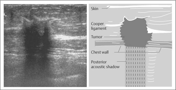
Multiple projections from the nodule within or around ducts extending away from the nipple usually seen in larger tumors. Shadowing no posterior acoustic features enhancement or combined pattern.

Posterior Acoustic Shadowing In Benign Breast Lesions Weinstein 2004 Journal Of Ultrasound In Medicine Wiley Online Library
It is wider than tall with macrolobulations no calcifications and posterior acoustic shadowing.

. As ultrasonic beams propagate through tissues there is a loss of energy by absorption reflection and scattering. Although posterior acoustic shadowing is a sonographic feature that is most commonly associated with mammary malignancies this sonographic finding may also be seen with benign breast lesions. Case Discussion Posterior acoustic shadowing and enhancement are two everyday key important concepts in ultrasound imaging.
Posterior acoustic features may or may not be seen when imaging solid masses. While breast US has certain advantages over digital mammography it suffers from image artifacts such as posterior acoustic shadowing PAS presence of which often obfuscates lesion margins. In this paper half-contour features are proposed to classify benign and malignant breast tumors with PAS considering the fact that the upper half of the tumor contour is less affected by PAS.
That posterior acoustic shadowing was more often. It is a form of imaging artifact. Posterior acoustic shadowing is a suspicious finding and may be seen in cases of invasive carcinoma postoperative scar complex sclerosing lesion or macrocalcifications and may even be seen in patients with dense breast tissue.
Citing Literature Volume 23 Issue 1 January 2004. 1 Because the ultrasonic transducer scans over the multiple tissue interfaces such as Cooper ligaments and other connective tissue posterior acoustic shadowing may result. D Magnified craniocaudal mammogram shows a spiculated mass arrow corresponding to the hypoechoic shadowing cancer.
A variety of pathologic conditions are discussed with pathologic-imaging correlation. While breast US has certain advantages over digital mammography it suffers from image artifacts such as posterior acoustic shadowing PAS presence of. Real-time sonographic evaluation of normal breast tissue may exhibit posterior acoustic shadowing.
The hepatic cyst demonstrates the opposite phenomenon of posterior acoustic enhancement. Posterior acoustic shadowing PAS can bias breast tumor segmentation and classification in ultrasound images. Which usually do not shadow.
This loss is displayed in the image as shadowing and is an important sonographic sign for the detection and diagnosis of breast disease. AbstractBreast ultrasound US in conjunction with digital mammography has come to be regarded as the gold standard for breast cancer diagnosis. While breast US has certain advantages over digital mammography it suffers from image artifacts such as posterior acoustic shadowing PAS presence of which often obfuscates lesion margins.
ILC may be occult in both mammography and ultrasound and breast-MRI may have certain advantages in the detection of this tumor type. Changes in Coopers ligaments. Posterior acoustic features.
Architectural distortion of the surrounding tissue. Transverse ultrasound of the left breast demonstrates an irregular antiparallel mass with posterior acoustic shadowing. At sonography cross section of a fat lobule can be mistaken for a solid mass that is isoechoic to surrounding adipose tis-.
Then the results from the systematic image interpretation were merged with the clinical data of the patients. Note also that the acoustic shadowing which is the dark black band posterior to the mass can be caused by productive fibrosis from the tumor and is often seen in masses that appear spiculated on the mammogram. Anyone have benign results with posterior acoustic shadowing brady0819 Upon a screening mammogram and ultrasound they found a 16 oval mass on my right breast.
What percentage of mammogram callbacks are cancer. It is the posterior acoustic shadowing that is freaking me out. The appearance of hazy echogenic material with posterior acoustic shadowing seen in an augmented breast should raise the suspicion for which of the following.
However on rescanning and dynamic imaging of the area particularly in a. The phenomenon of acoustic shadowing sometimes somewhat tautologically called posterior acoustic shadowing on an ultrasound image is characterized by a signal void behind structures that strongly absorb or reflect ultrasonic waves. Posterior acoustic shadowing is suspicious for breast cancer If a breast lesion shows posterior acoustic shadowing on ultrasound this means that there is something about the mass or around the mass which attenuates reduces the sonic beam strength in comparison to normal adjacent tissues.
Fat Lobule The normal breast is composed of numer- ous fat lobules mixed with dense fibroglan- dular tissue. On mammography the lesion usually shows localized increased density in the glandular tissue. Sonographic posterior acoustic shadowing.
DMP usually shows nonspecific parenchymal enhancement rather than an irregular enhancing mass on MRI. Ultrasound Longitudinal The calculi in the kidney and gallbladder cause pronounced posterior acoustic shadowing. An irregular hypoechoic mass with intense posterior acoustic shadowing can be typically seen on US and can mimic breast malignancy Fig.
The intense posterior acoustic shadow pro- duced by the whirled smooth muscle bundles of the nipple. Shadowing may result because of reflection of most of the energy by a large impedance discontinuity. Sonographic Appearance of Invasive Ductal Carcinoma of the Breast According to Histologic Grade W402 AJR199 September 2012 ulated mass margins and acoustic shadowing they found that higher-grade tumors were sig - nificantly more likely than lower-grade ones to display poorly defined margins and posteri - or acoustic enhancement.
Breast ultrasound US in conjunction with digital mammography has come to be regarded as the gold standard for breast cancer diagnosis.

Pdf Distinguishing Lesions From Posterior Acoustic Shadowing In Breast Ultrasound Via Non Linear Dimensionality Reduction Semantic Scholar

Posterior Acoustic Shadowing In Benign Breast Lesions Weinstein 2004 Journal Of Ultrasound In Medicine Wiley Online Library

Basic Principles Radiology Key

Posterior Acoustic Shadowing In Benign Breast Lesions Weinstein 2004 Journal Of Ultrasound In Medicine Wiley Online Library

Mediconotebook Posterior Acoustic Shadowing And Enhancement

Ultrasound Image Of A Breast Cancer With Irregular Borders Angular Download Scientific Diagram

Transverse Ultrasound Of The Left Breast Demonstrates An Irregular Download Scientific Diagram
0 comments
Post a Comment